Table Of Content

It is important to note that while a 3D ultrasound is a reliable way to determine the gender of a baby, it is not always 100% accurate. In some cases, the sonographer may mistake the genital area for something else, or the baby’s position may make it hard to see their genital area clearly. 5D ultrasound show the realistic view or what many call the flesh tone look of baby. 2D is like your good old reliable car, often covered by insurance and kinder to the wallet. But hey, it’s a once-in-a-lifetime movie ticket to see your baby, so weigh those pros and cons.
My Baby Has Hair! What Does That Mean? 🧐
Hair can appear as either a bright white or dark black line depending on the type of hair and its thickness. However, in some cases, it may be difficult to differentiate between a hair and other structures in the body due to their similar densities. Additionally, it is important to note that ultrasounds cannot detect individual strands of hair but rather only larger clumps of hair follicles.
Moms Share Home Remedies for Pregnancy Morning Sickness
You’re more likely to see hair towards the end of your pregnancy than at the start or middle. Your insurance might not cover them, and your doctor may steer you away from getting them for nonmedical reasons. Ultrasound can detect the presence of an abnormality and determine its location and severity. Benign cysts are not cancerous and do not spread to other parts of the body. Ultrasound is often used to diagnose and monitor the progress of these abnormalities. While this halo can make it difficult to see fine details, it can also provide important information about the overall shape and size of objects.
If my baby is hairy in the womb, will that cause me heartburn?
They produce a more realistic image of a baby but do not capture hair strands, and it is in this regard, technically, advancement in this ultrasound did not mean the 2D ultrasound is lesser. It uses sound waves with pictures taken at different angles to create a 3D view. Since the image is in black and white, the hair appears like a fuzz on the head of the baby, and while it may not be noticeable to a layperson, trained professionals can point it out.
Baby girl's hair showed up on her ultrasound scan - Daily Mail
Baby girl's hair showed up on her ultrasound scan.
Posted: Thu, 16 Feb 2017 08:00:00 GMT [source]

3D ultrasounds are a type of medical imaging technique that uses sound waves to create three-dimensional images of the developing fetus. The purpose of a 3D ultrasound is to provide a more detailed view of the baby’s facial features, organs, and overall development than traditional 2D ultrasounds. 5D ultrasound is commonly used during pregnancy to monitor fetal development and detect any potential health issues.
When Hair Becomes Visible on Ultrasound
While the exact reason behind this isn’t known, it’s another example of how genetics influence the amount of hair a baby is born with. Experts aren’t entirely sure why only some babies are born with a full head of hair, but genetics and hormones are thought to play a significant role. The hair that grows in afterward, called terminal hair, is often a different color, thickness, and texture than the hair the baby was born with.
While ultrasounds give us a sneak peek, they’re just part of the prenatal bonding experience. Talking to your baby, playing music, and imagining the future are all ways parents connect with their unborn child, hair unseen. But remember, it’s more about the conditions during the ultrasound than the type itself. Some parents mistake lack of hair in ultrasound images and the first few months of a child’s birth as baldness. There are a variety of factors that ultimately determine if we are able to see hair in a 3D ultrasound. Many expecting parents are curious if they will be able to see the baby’s hair during their 3D ultrasound session.

Hair is typically visible on a 3D ultrasound after 25 weeks of gestation. Overall, a 5D ultrasound is a safe and non-invasive procedure that allows healthcare professionals and parents to obtain a more detailed and lifelike view of the developing fetus in the womb. It’s also important to note that 3D ultrasounds are typically not covered by insurance and can be expensive.
It can also be used for gender determination and to create keepsake images and videos for parents. However, it is important to note that 5D ultrasound is not a replacement for medical ultrasound exams and should only be performed by trained healthcare professionals. The images are black and white and show the structure and shape of the object being scanned. 2D ultrasounds are used for routine prenatal care, as well as to diagnose and monitor medical conditions. During pregnancy, 3D ultrasounds can be used to monitor fetal growth and development, detect abnormalities or birth defects, and determine the gender of the baby.
The reason in utero hair (lanugo) develops is to keep the baby warm, and around 30 to 32 weeks of gestation, just eight weeks away from your due date, your baby can lose that hair. This is because they’re gaining body fat, so they no longer need that wispy, downy soft lanugo. If you or your partner have a head full of hair, chances are your baby will too. My last ultrasound was a 3D one, and it’s amazing how they can detect lots of hair on a fetus. A 3D ultrasound creates a still image of the baby, while a 4D ultrasound creates a moving image.
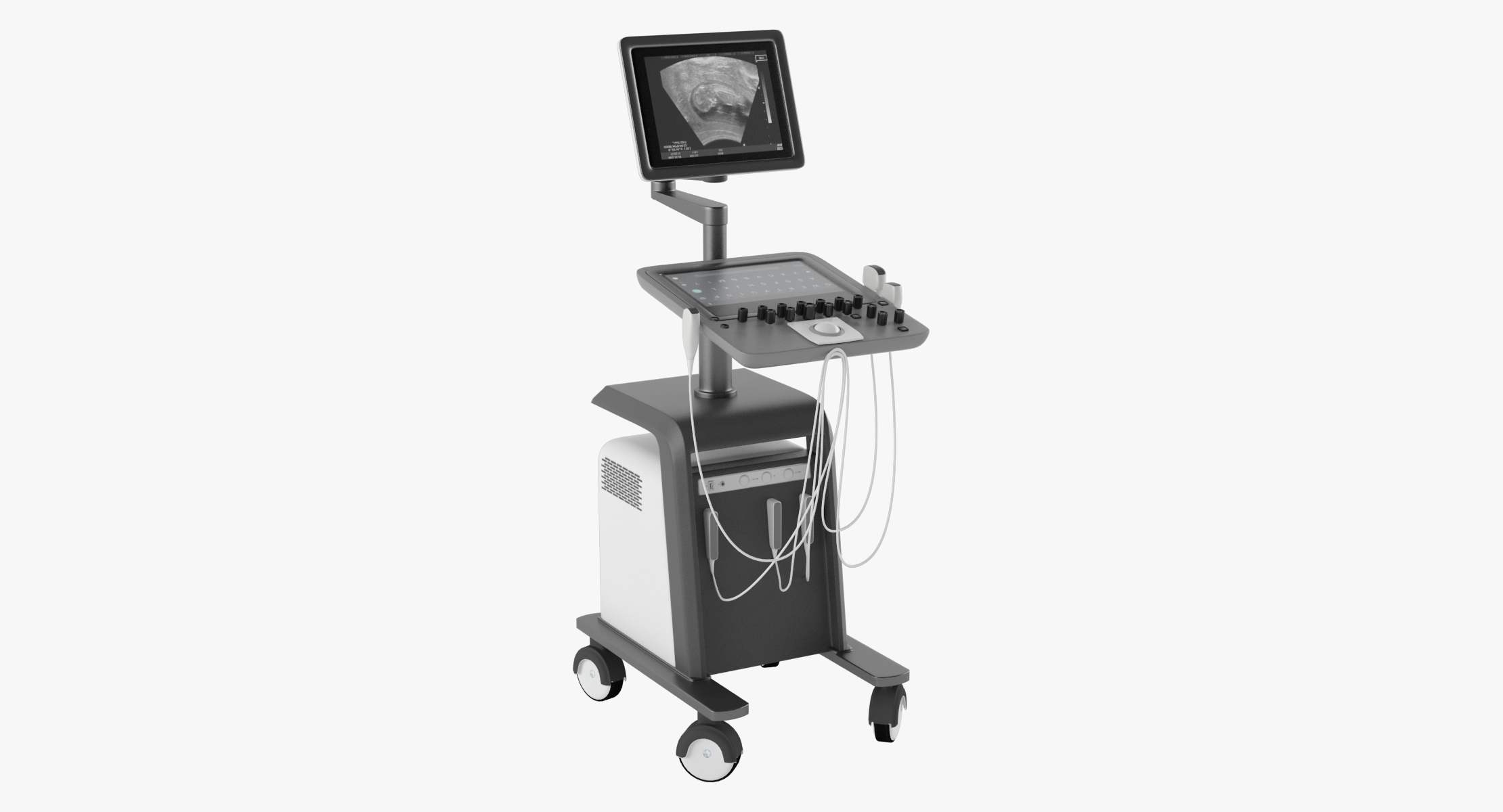
If you do see hair in a late ultrasound, it will probably look like white strands on the scalp, or a fuzzy white halo. It is important to note that keepsake ultrasounds should not be used as a substitute for medical ultrasounds. While 3D/4D images can be a fun way to connect with the baby, they should not be relied upon for medical information or used to diagnose any potential complications. The ultrasound probe, also known as a transducer, is then moved back and forth over the skin to capture images of the fetus. The probe emits high-frequency sound waves that bounce off the fetus and surrounding tissues, creating images that can be viewed on a monitor. The appearance of hair on a 3D or 4D ultrasound can provide an exciting glimpse into what the baby will look like after it’s born.
Lanugo is a very fine, soft, and downy type of hair that covers the body of a developing fetus. Lanugo and true baby hair are two different types of hair that infants can have during different stages of development. The second ultrasound, between 18 and 22 weeks, is to check the fetal anatomy for abnormalities, infections, and growth. An old wives’ tale says that heartburn during pregnancy indicates that your baby will have a lot of hair.
The fetus also develops hair on other parts of the body, such as the back, arms, and legs. Overall, while it is possible to see hair on a 3D ultrasound, the visibility of hair strands and follicles can vary depending on factors such as hair thickness, color, and growth. It is also important to note that not all babies have hair visible on a 3D ultrasound. Hair growth can vary from baby to baby, and some babies may not have enough hair to be visible on the ultrasound. It also allows parents to see their baby’s features and movements in greater detail, which can be a very emotional and rewarding experience. Ultrasounds are an essential medical technology because they allow external examination and diagnosis.
Due to its small size and light color, fine baby hair may not be visible until later in the pregnancy when it has grown thicker and darker. It’s important to note that while 3D ultrasounds can be a fun and exciting way to connect with your baby before birth, they are not a medical necessity. Traditional 2D ultrasounds are still the go-to for prenatal check-ups and monitoring your baby’s development. Ultimately, the decision to have a 3D ultrasound comes down to personal preference, and it’s always wise to discuss your options with your healthcare provider. It takes multiple three-dimensional images in real time, creating a moving video of your baby in the womb.
During the appointment, the patient will be asked to lie down on a table and a special gel will be applied to the skin over the area that will be examined. Overall, the decision to get a 3D ultrasound is a personal one and should be discussed with a healthcare provider. It is important to choose a reputable facility that uses safe and reliable equipment, and to follow any instructions or precautions provided by the healthcare provider. 3D ultrasounds are often used to diagnose fetal abnormalities and to monitor fetal growth and development. It can be tempting to want to get a sneak peek at your developing baby at all stages of growth, but what week is best for a 3D ultrasound?

No comments:
Post a Comment